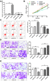
hsa-miR-4443 inhibits myocardial fibroblast proliferation by targeting THBS1 to regulate TGF-β1/α-SMA/collagen signaling in atrial fibrillation
- PMID: 33681892
- PMCID: PMC7931814
- DOI: 10.1590/1414-431X202010692
hsa-miR-4443 inhibits myocardial fibroblast proliferation by targeting THBS1 to regulate TGF-β1/α-SMA/collagen signaling in atrial fibrillation
Abstract
Fibrosis caused by the increase in extracellular matrix in cardiac fibroblasts plays an important role in the occurrence and development of atrial fibrillation (AF). The aim of this study was to investigate the role of hsa-miR-4443 in AF, human cardiac fibroblast (HCFB) proliferation, and extracellular matrix remodeling. TaqMan Stem-loop miRNA assay was used to measure hsa-miR-4443 expression in patients with persistent AF (n=123) and healthy controls (n=100). Patients with AF were confirmed to have atrial fibrosis by late gadolinium enhancement. At the cellular level, after hsa-miR-4443 mimic and inhibitor were transfected with HCFBs, proliferation, apoptosis, migration, and invasion were analyzed. Lastly, hsa-miR-4443-targeted gene and transforming growth factor (TGF)-β1/α-SMA/collagen pathway were evaluated by dual-luciferase reporter assay and western blot, respectively. In patients with AF, hsa-miR-4443 decreased significantly and collagen metabolism level increased significantly. Logistic regression analysis showed that low hsa-miR-4443 level was a risk factor of AF (P<0.001). The receiver operating characteristic curve revealed that hsa-miR-4443 was useful for predicting AF (area under the curve: 0.828, sensitivity: 0.71, specificity: 0.78, P<0.001). In HCFBs, hsa-miR-4443 targeted thrombospondin-1 (THBS1) and downregulated TGF-β1/α-SMA/collagen pathway. The inhibition of hsa-miR-4443 expression promoted HCFB proliferation, migration, invasion, myofibroblast differentiation, and collagen production. The significant reduction of hsa-miR-4443 can be used as a biomarker for AF. hsa-miR-4443 protected AF by targeting THBS1 and regulated TGF-β1/α-SMA/collagen pathway to inhibit HCFB proliferation and collagen synthesis.
Figures


Similar articles
-
Modulation of miR-10a-mediated TGF-β1/Smads signaling affects atrial fibrillation-induced cardiac fibrosis and cardiac fibroblast proliferation.Biosci Rep. 2019 Feb 8;39(2):BSR20181931. doi: 10.1042/BSR20181931. Print 2019 Feb 28. Biosci Rep. 2019. PMID: 30683806 Free PMC article.
-
MiRNA-1202 promotes the TGF-β1-induced proliferation, differentiation and collagen production of cardiac fibroblasts by targeting nNOS.PLoS One. 2021 Aug 24;16(8):e0256066. doi: 10.1371/journal.pone.0256066. eCollection 2021. PLoS One. 2021. PMID: 34428251 Free PMC article.
-
Quercetin improves atrial fibrillation through inhibiting TGF-β/Smads pathway via promoting MiR-135b expression.Phytomedicine. 2021 Dec;93:153774. doi: 10.1016/j.phymed.2021.153774. Epub 2021 Sep 26. Phytomedicine. 2021. PMID: 34656066
-
MicroRNA-21 via Dysregulation of WW Domain-Containing Protein 1 Regulate Atrial Fibrosis in Atrial Fibrillation.Heart Lung Circ. 2018 Jan;27(1):104-113. doi: 10.1016/j.hlc.2016.01.022. Epub 2017 Mar 12. Heart Lung Circ. 2018. PMID: 28495464
-
Rapid atrial pacing induces myocardial fibrosis by down-regulating Smad7 via microRNA-21 in rabbit.Heart Vessels. 2016 Oct;31(10):1696-708. doi: 10.1007/s00380-016-0808-z. Epub 2016 Mar 11. Heart Vessels. 2016. PMID: 26968995 Free PMC article.
Cited by
-
Epigenetic Modification Factors and microRNAs Network Associated with Differentiation of Embryonic Stem Cells and Induced Pluripotent Stem Cells toward Cardiomyocytes: A Review.Life (Basel). 2023 Feb 17;13(2):569. doi: 10.3390/life13020569. Life (Basel). 2023. PMID: 36836926 Free PMC article. Review.
-
Circulating MicroRNAs as Specific Biomarkers in Atrial Fibrillation: A Meta-Analysis.Noncoding RNA. 2023 Feb 9;9(1):13. doi: 10.3390/ncrna9010013. Noncoding RNA. 2023. PMID: 36827546 Free PMC article. Review.
-
Aberrant expression of microRNA-4443 (miR-4443) in human diseases.Bioengineered. 2022 Jun;13(6):14770-14779. doi: 10.1080/21655979.2022.2109807. Bioengineered. 2022. PMID: 36250718 Free PMC article. Review.
-
Association of THBS1 genetic variants and mRNA expression with the risks of ischemic stroke and long-term death after stroke.Front Aging Neurosci. 2022 Sep 23;14:1006473. doi: 10.3389/fnagi.2022.1006473. eCollection 2022. Front Aging Neurosci. 2022. PMID: 36212039 Free PMC article.
-
Mining Potential Drug Targets and Constructing Diagnostic Models for Heart Failure Based on miRNA-mRNA Networks.Mediators Inflamm. 2022 Sep 27;2022:9652169. doi: 10.1155/2022/9652169. eCollection 2022. Mediators Inflamm. 2022. PMID: 36204659 Free PMC article.
References
MeSH terms
Substances
LinkOut - more resources
Full Text Sources
Other Literature Sources
Medical
Miscellaneous

