. 2020 Jan;18(1):105-117.
doi: 10.1158/1541-7786.MCR-19-0239. Epub 2019 Oct 18.

Targeting the Kynurenine Pathway for the Treatment of Cisplatin-Resistant Lung Cancer

Affiliations
Free PMC article

Targeting the Kynurenine Pathway for the Treatment of Cisplatin-Resistant Lung Cancer

Dan J M Nguyen et al. Mol Cancer Res. 2020 Jan.
Free PMC article

Abstract

Cisplatin resistance is a major barrier in the effective treatment of lung cancer. Cisplatin-resistant (CR) lung cancer cells do not primarily use glucose but rather consume amino acids such as glutamine and tryptophan (Trp) for survival. CR cells activate the kynurenine (KYN) pathway (KP) to cope with excessive reactive oxygen species (ROS) and maintain homeostasis for growth and proliferation. Consequently, indoleamine 2,3-dioxygenase-1 (IDO1) becomes an essential enzyme for CR cells' survival because it initiates and regulates the first step in the KP. Increased IDO1 activities and ROS levels are found in CR cells versus cisplatin-sensitive lung cancer. Importantly, significantly greater KYN/Trp ratio (P = 0.005) is detected in serum of patients who fail cisplatin when compared with naïve treatment. Knocking down IDO1 using shRNA or IDO1 inhibitors heightens ROS levels and results in a significant growth inhibitory effect only on CR cells and not on cisplatin-sensitive cells. Exposing CR cells to antioxidant (TIRON) results in suppression of IDO1 activity and confers resistance to IDO1 inhibition, indicating an interrelationship between ROS and IDO1. Because KYN plays a critical role in reprogramming naïve T cells to the immune-suppressive regulatory T-cell (T-reg) phenotype, we observed higher expression of TGFβ, FoxP3, and CD4+CD25+ in mice bearing CR tumors compared with tumors from cisplatin-sensitive counterparts. IMPLICATIONS: Findings suggest that the enzyme-inhibitory activity and antitumor efficacy of IDO1 inhibitors rely in part on ROS levels, arguing that IDO1 expression alone may be insufficient to determine the clinical benefits for this class of experimental cancer drugs. Importantly, IDO1 inhibitors may be more suitable to treat patients with lung cancer who failed cisplatin therapy than naïve treatment patients.

Conflict of interest statement

DISCLOSURE OF POTENTIAL CONFLICTS OF INTEREST

No potential conflicts of interest were disclosed.

Figures

Figure 1.
Figure 1.
CR lung cancer cells relied on OXMET and utilized KP which contributed to the production of kynurenine (KYN) in CR tumors. (A) Parent (A and FA) and CR (ALC and FC) cells were assayed for baseline oxygen consumption using Seahorse XFe24 extracellular flux analyzer. The rate of oxygen consumption (OCR) was 6 times higher in CR than parental cells (*p=0.009 and **P=0.003). LL24 was used as control. (B) Tryptophan (TRP) uptake was determined in parental vs CR cells using L-[5-3H(N)] tryptophan. The rate of TRP uptake was higher in CR cells (*p=0.02, **p=0.007). (C) Total cellular NAD+ concentrations were detected in parental vs. CR cell lines. CR possessed lower basal levels of NAD+ (*p=0.02; **p=0.006). (D) Relative mRNA levels of QPRTase. QPRTase mRNA was significantly decreased in CR cells. GAPDH was used as internal control. The results shown in the graph were calculated with the ΔΔCt method by setting the mRNA level of parental cells as 1 (*p=0.04, **p=0.01). (E) Extracellular TRP concentration in culture medium were determined by amino acid analyzer. Lower levels of TRP were found in CR cells’ culture media (*p=0.04, **p=0.02). (F) Extracellular KYN concentration in culture medium were determined by amino acid analyzer. Higher levels of KYN were found in CR cells’ culture media (*p=0.01, **p=0.005). (G) Extracellular KYN/TRP ratio in culture medium were determined by HPLC. CR cells possessed significantly greater KYN/TRP ratio (*p=0.003, **p=0.01) than control. (H) KYN/TRP ratio in patients’ serum (HPLC). Patients 1–3 failed cisplatin after third cycle of cisplatin (*p=0.002, **p=0.004, ***p=0.005). Patients 4–6 continued to respond to cisplatin after the third cycle of treatment. (Note: Pt = patient).
Figure 2.
Figure 2.
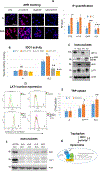
Modulator role of KYN/AHR/ARNT axis in CR cells metabolism. (A) Immunofluorescence (IF) staining of cells with 1:1000 AHR antibody (red) and DAPI (blue nuclei). CR cells (ALC) possessed significantly higher intensity of AHR expression in the nucleus when compared to parental cells (*p=0.008). DMF or CH223191 significantly reduced AHR accumulation in CR cells (**p=0.007, ***p=0.001; respectively) while exposure to KYN markedly increased AHR accumulation in the nucleus of both parental and CR cells. Bar graph indicated quantification of IF intensity (RFU/cell) using hybrid cell count. (B) Addition of 100μM of KYN increased IDO1 activity in CR cells substantially (*p=0.03), but not significantly (NS) in parental cells. Treatment of 10μM of DMF or 1μM of CH223191 resulted in significant suppression of IDO1 activities (**p=0.006, ***p=0.002, respectively). (C) Immunoblot of lung cancer cell lines showed that resistant variants did not possess HIF1α, but expressed higher levels of AHR and LAT1. Actin was used as a loading control. (D) Flow cytometry analysis of surface LAT1 in lung cancer cell lines. D1:CR cells possessed higher surface LAT1 when compared to parental counterparts. D2:Treatment of KYN at 100μM for 48h further enhanced LAT1 expression in CR cells. D3:KYN treatment did not increase LAT1 expression in parental cells. D4:Knocking down HIF1α in parental cells resulted in increased LAT1 expression after KYN treatment. (E) Knocking down HIF1α in parental cells increased TRP uptake and further increased TRP uptake upon exposure to KYN (100μM). Treatment of KYN further heightened TRP uptake in CR cells (48h; *p=0.02, **p=0.006). (F) Knocking down IDO1 (shIDO1) in ALC suppressed LAT1 expression. Sh-A to D represent 4 unique shRNA sequences. (G) Diagram illustrating the binding partners of ARNT (HIF1β). When HIF1α is down-regulated, ARNT formed a new binding partner with AHR/KYN and initiated the transcription of genes that favored proliferation and increased TRP uptake which can lead to further KYN secretion in CR cells.
Figure 3.
Figure 3.
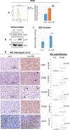
Increase IDO1 activity and immune suppressive phenotype were found in CR tumor. (A) ROS analysis of mouse (LLC vs LLC-CR) cells. LLC-CR expressed 2 fold higher basal level of ROS. (B) Immunoblot showed that LLC-CR also expressed higher levels of LAT1 protein. (C) LLC-CR possessed higher IDO1 activity (*p=0.04). (D) Immunohistochemistry (IHC) staining of intratumoral CD4+, CD25+, FoxP3+ (arrow), TGFβ, and IDO1. Higher T-reg densities were found in mice bearing CR tumor than control. Box graph indicated quantification of IHC (intensity/μM2) using hybrid cell count.
Figure 4.
Figure 4.
IDO1 expression alone is not the only factor in determining its activity. (A) Diagram illustrated that tryptophan can be used to generate serotonin and KYN. (B) Relative mRNA levels of IDO1. Total RNAs extracted from these cells were reverse-transcribed and subsequently used as template for real-time quantitative PCR. GAPDH was used as internal control. The results shown in the graph were calculated with the ΔΔCt method by setting the IDO1 mRNA level of LL24 cells as 1 (*p=0.005, **p=0.01). (C) Immunoblot of lung cancer cell lines treated with and without IFNγ (20ng/ml) for 24h. No significant differences in IDO1 protein expression levels were observed between parental and CR cells. (D) Significantly higher IDO1 activity was found in CR cells when compared to parental cell counterparts (*p=0.02, **p=0.03) and was further enhanced with IFNγ (20ng/ml) treatment for 24h (***p=0.03, ****p=0.008).
Figure 5.
Figure 5.
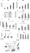
Antitumor activity of IDO inhibitors in CR cells. (A) Growth inhibitory effect of various IDO inhibitors (Epacadostat: EPA, NLG-919: NLG, PF-06840003: PF, or Indoximod: INDO) for 48hrs showed that CR cells were sensitive to IDO1 inhibitors with EPA yielding the best efficacy (*p=0.03, **p=0.02). (B) ID50 dosage of EPA alone and in combination with 20ng/ml of IFNγ. Combination treatment enhance EPA toxicity only in CR cells. (Mean SD of three experiments, 48h). (C) ROS analysis detected by CM-H2DCFDA probe indicated that CR cells expressed higher basal levels of ROS. (D) ROS levels were heightened when treated with 15μM of EPA for 48hrs (*p=0.009, **p=0.005). Bar graph represents the relative fluorescent units/cell via fluorometer plate reader. (E) Immunoblot of lung cancer cell lines treated with and without EPA (15μM/ml) for 48h.
Figure 6.
Figure 6.
Sensitivity to IDO1 inhibitor is dependent with higher ROS levels. (A) ID50 dosage of cisplatin in parental cell line “A” and its cisplatin resistance variants. (B) Intracellular ROS production measured by fluorescence intensity using CM-H2DCFDA probe. (C) ID50 dosage of EPA and its cisplatin resistance variants. CR2, CR4, and CR6 possessed 2, 4, and 6 fold resistance to cisplatin, respectively (Mean SD of three experiments, 48h). (D) Knocking down of IDO1 (shIDO-A and shIDO-B) further enhanced ROS level in CR cells (*p=0.02,**p=0.03); however, treatment with 15μM of EPA for 48hrs did not produce additional ROS accumulation in ALCshIDO. (E) Growth inhibitory effect of EPA (48hrs) on ALC and ALCshIDO. EPA significantly suppressed ALC cells’ growth (*P=0.002), and no significant growth inhibition were observed in the knocked down clones. (F) Intracellular ROS production. Antioxidant (TIRON) suppressed ROS production in CR cells (*p=0.003, **p=0.006). (G) Treatment of TIRON (48hrs) attenuated IDO1 activity (*p=0.03) in CR cells. (H) TIRON treatment conferred resistance to EPA in CR cells (48h; **p=0.02). (I) CR cells do not primarily utilize glucose, but rather consume amino acids such as glutamine and tryptophan for survival. This metabolic switch is due to increased ROS production hyper-activating kynurenine pathway (KP) to balance oxidative stress and maintain cellular growth and proliferation. Kynurenine (KYN) is oxidized through indoleamine 2,3-dioxygenase (IDO), and plays a key role in reprogramming naïve T-cells to the immune suppressive regulatory T-cell (T-reg) phenotype. Further increase in ROS by interfering with tumor metabolism via IDO1 inhibiting will selectively target these cisplatin resistant lung cancer cells.

Similar articles

Cited by 13 articles

Publication types

MeSH terms